Submitted by Sarah Faulwetter on Fri, 2012-04-06 16:26
Gallery:
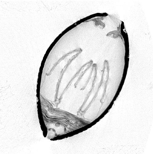
MicroCT images Bivalve, cross section, ca. 2mm. Sample has been stained with phosphotungstic acid before scanning to enhance contrast. Muscle fibres and gills are clearly visible. Scanned with a SkyScan 1172 micro-CT.trace affiliate link | Nike Shoes